Microscopy
TEM Images
Transmission electron microscopy was pioneered by Ernst Ruska and Max Knoll in 1931 — work that earned Ruska the Nobel Prize in Physics over five decades later. Rather than light, TEM fires a beam of electrons through an ultrathin specimen; because electrons have wavelengths thousands of times shorter than visible photons, the technique resolves features down to the ångström scale, far below the diffraction limit of any optical instrument. The micrographs below use negative staining, in which a heavy-metal salt such as uranyl acetate floods the background and is repelled by the nanostructure, rendering the surrounding field dark and the structure bright. Each image is paired with an interactive 3D model of the corresponding self-assembled nanostructure.
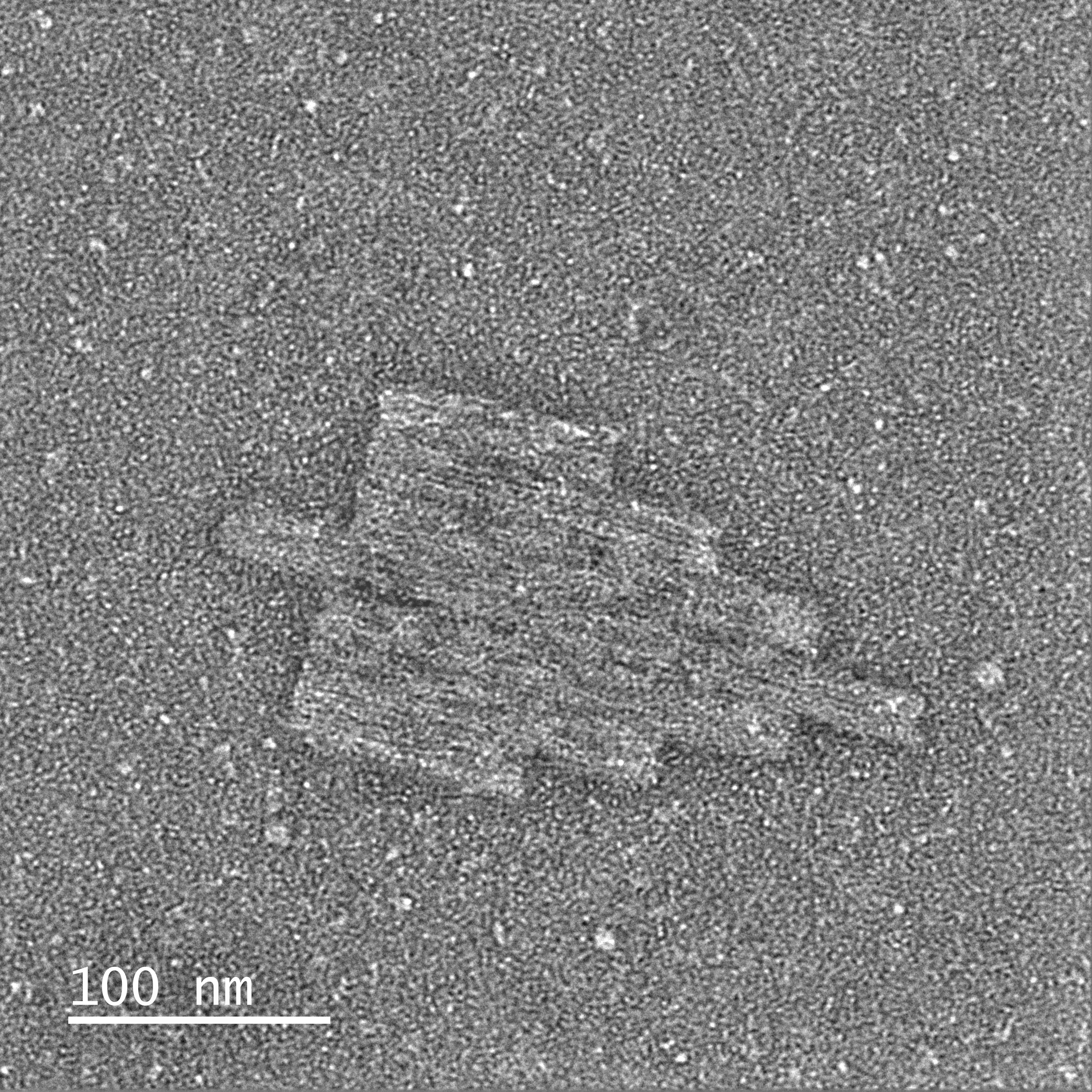
Peace
Negatively-stained TEM micrograph of the Peace nanostructure at ×80,000 magnification.Crown
Negatively-stained TEM micrograph of the Crown nanostructure at ×80,000 magnification.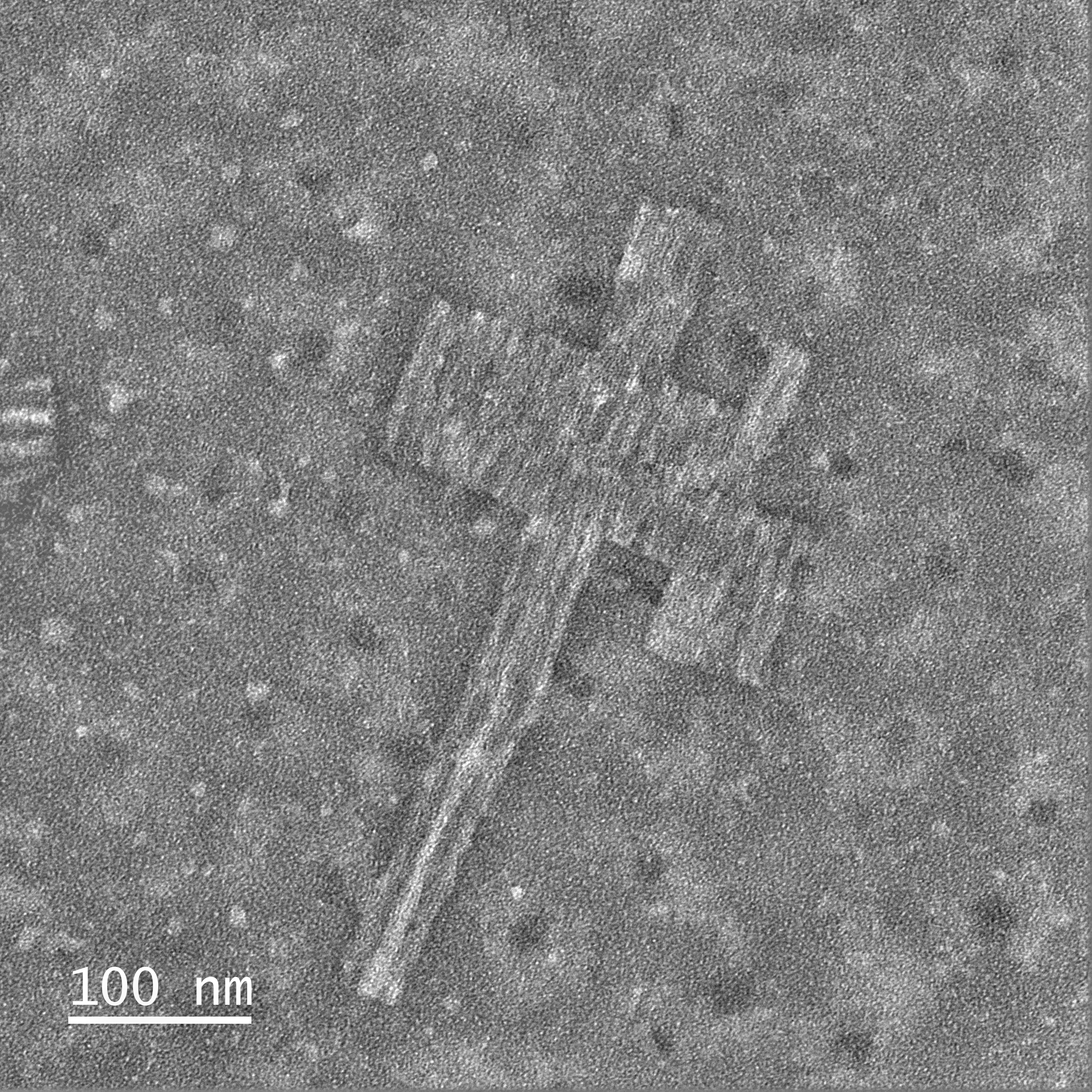
Cross
Negatively-stained TEM micrograph of the Cross nanostructure at ×80,000 magnification.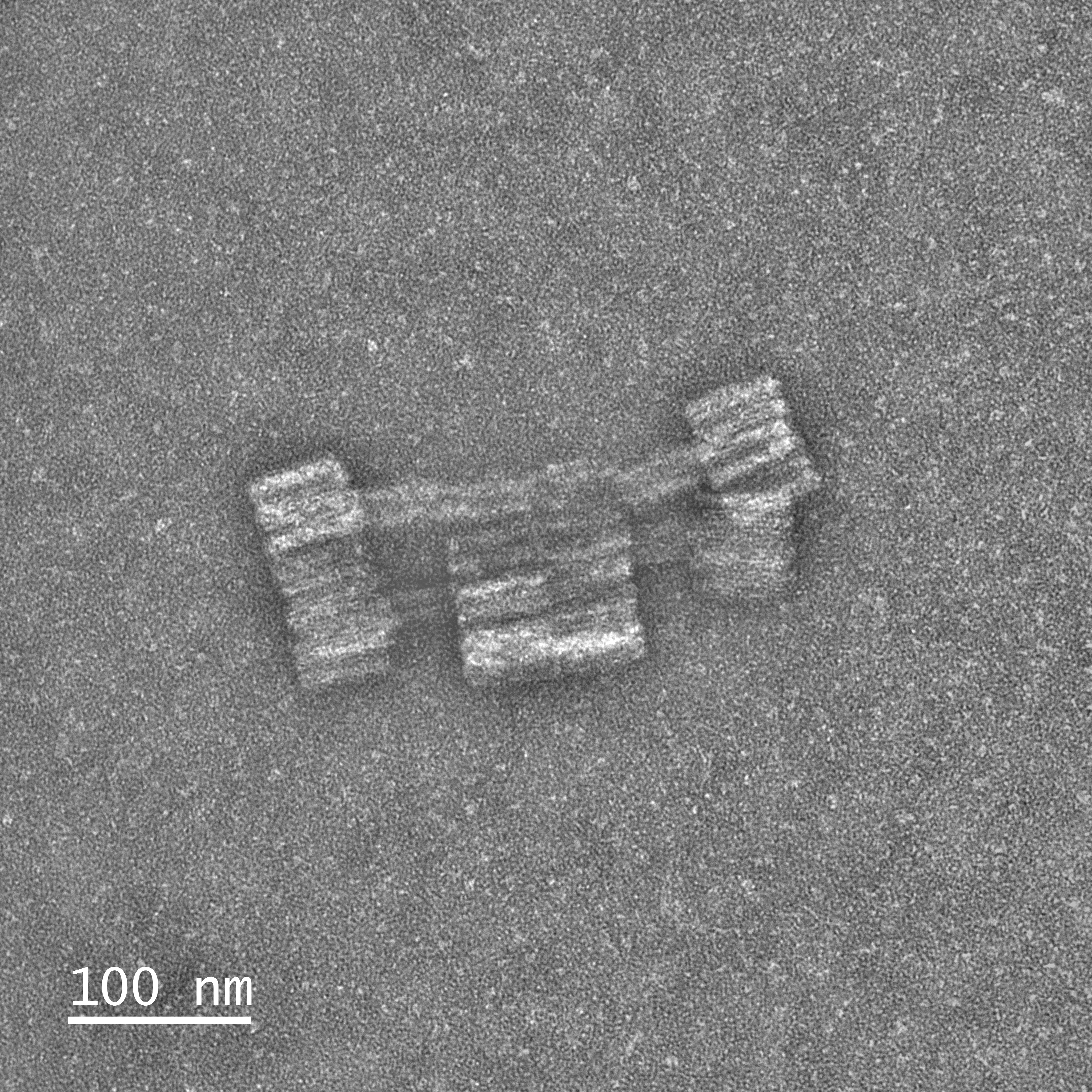
Mars Rover
Negatively-stained TEM micrograph of the Mars Rover nanostructure at ×80,000 magnification.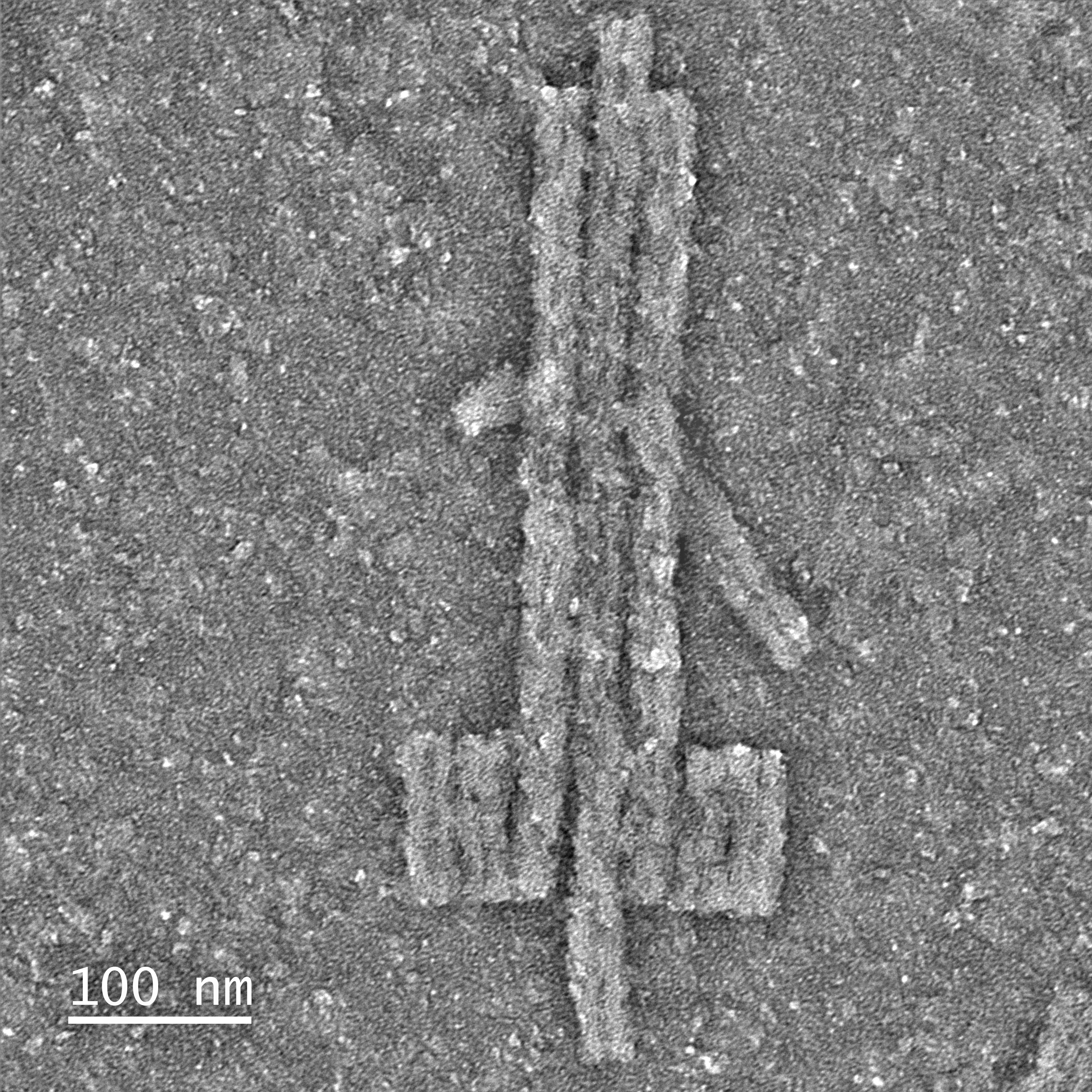
Sword
Negatively-stained TEM micrograph of the Sword nanostructure at ×80,000 magnification.